The Depth of Pigmentation: Why Location Changes Everything
Two dark patches can look identical in the mirror and be completely different diagnoses. What separates them is not colour or size — it is how deep the pigment sits.
 Dr. Jihye Kim
Medical doctor, specialist, Laplace Clinic · Laplace Clinic, Sinchon, Seoul · July 28, 2026 · 7 min read
A patient points to two marks on her cheek. They are the same colour, roughly the same size, and appeared within a year of each other. She assumes they are the same problem. In the clinic they are two different diagnoses with two different treatment plans and two very different timelines — and the only thing that separates them is depth.
Pigment is the area of dermatology where patient expectations and clinical reality diverge the most. Almost always, the gap comes from the same misunderstanding: that pigmentation is one condition, graded by how dark it looks. It is not. It is a group of conditions sorted by where in the skin the pigment is sitting.
Three Layers, One Colour
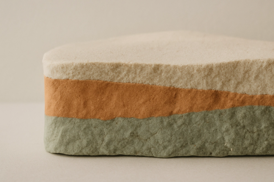
It helps to picture the skin as three broad zones. The epidermis is the outermost layer — thin, constantly renewing itself, and the layer you touch. Beneath it is the dermis, thicker and more stable, holding the collagen, vessels and structure that give skin its resilience. Below that is the subcutaneous layer.
Melanin can end up in the epidermis, in the dermis, or in both. To the eye these can look similar, because you are seeing the same pigment through different amounts of overlying tissue. To the clinician they are separate problems, because the epidermis renews and the dermis does not — at least not on any timescale a patient would recognise.
What Sits in the Epidermis
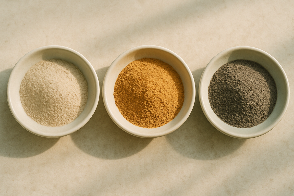
Superficial pigment includes the marks most people arrive worried about: sun-induced spots and lentigines, freckles, general uneven tone from cumulative sun exposure, and much of the brown discolouration left behind after acne — post-inflammatory hyperpigmentation.
Epidermal pigment tends to look sharply defined, sits on the brown side of the spectrum, and often becomes more obvious under direct light. Its great advantage is location. It is in a layer that is designed to turn over, which means treatment can work with the skin's own renewal rather than against it.
What Sits in the Dermis
Deeper pigment behaves differently. It includes dermal melanocytic conditions such as nevus of Ota and related birthmark-type pigmentation, some persistent grey-brown patches, tattoo ink, and the deeper component of long-standing melasma.
Seen through the epidermis, dermal pigment often reads as greyer, bluer, or hazier than superficial pigment, and its borders are usually less distinct. It also does not respond to surface renewal, because there is no renewal happening at that depth. Whatever treats it has to reach it, and then the body has to physically remove what is left.
Epidermal pigment is a layer problem. Dermal pigment is a transport problem.

Why Melasma Is a Category of Its Own
Melasma deserves a separate paragraph because it is the most common reason a pigment plan goes wrong. It is frequently mixed — partly epidermal, partly dermal — and it is driven by factors that no laser addresses: hormonal influence, heat, visible light as well as ultraviolet, and an underlying tendency of the melanocytes to over-respond to stimulation.
The clinical consequence is counter-intuitive. With melasma, treating harder frequently makes it worse. Aggressive energy delivered to skin that is already inflamed and reactive can trigger a rebound that leaves the patient darker than when they started. This is why an experienced clinician may recommend a cautious, maintenance-oriented plan for melasma while confidently clearing a sun spot on the same face in a single session.
How Depth Is Assessed
Depth is judged clinically, not guessed. The assessment usually combines several things: the colour and border quality of the lesion, its history — how long it has been present, how it began, whether it fluctuates with season or hormones — the response of the surrounding skin, and examination under controlled lighting and magnification, sometimes with imaging that visualises pigment beneath the surface.
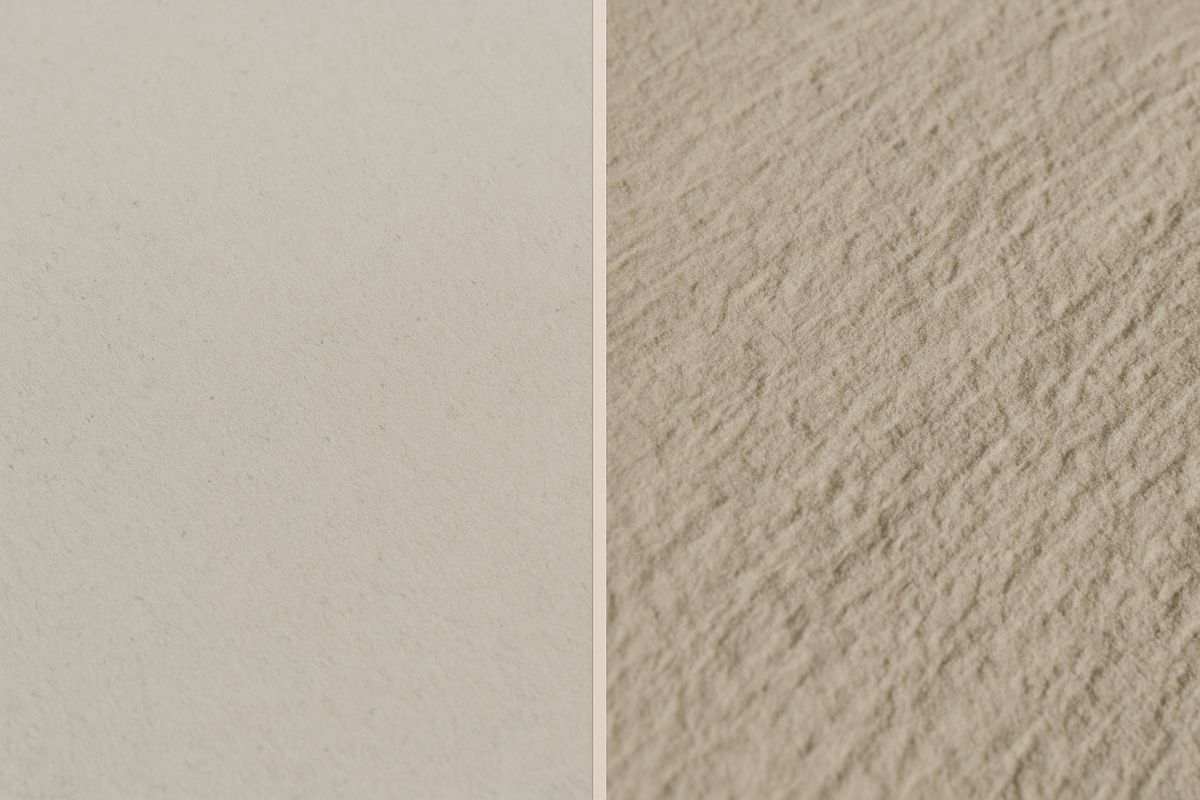
Which is also why a diagnosis by photograph is unreliable. A phone image compresses exactly the information that distinguishes surface pigment from deep pigment: subtle colour temperature, border definition, and the way the mark behaves under different light.
What This Means for You
A few practical conclusions follow from depth, and they are worth knowing before your first consultation.
- A single treatment plan cannot cover every dark mark on your face. Different depths on the same cheek may be treated on entirely different schedules.
- "How many sessions" has no general answer. It is a function of depth, not of severity — a faint but deep patch may take far longer than a dark but superficial one.
- Faster is not better. Pigment treated too aggressively can rebound, particularly in melasma and in skin prone to post-inflammatory darkening.
- Sun protection is not an accessory to pigment treatment. It is the treatment's operating condition — without it, you are removing pigment while continuing to produce it.
The most useful question you can ask at a consultation is not which laser will be used. It is: how deep is this, and what does that mean for how long this will take? A clinic that can answer specifically has made a diagnosis. In the next article, I look at what actually happens to pigment once it is treated — and why the two depths clear through completely different biological routes.
Frequently Asked Questions
How do I know if my dark spots are superficial or deep?
You generally cannot tell reliably at home. Superficial pigment tends to be brown with defined borders; deeper pigment often looks greyer or hazier with indistinct edges. Accurate assessment uses history, controlled lighting, magnification and sometimes imaging that visualises pigment below the surface.
Why does my friend's pigmentation clear faster than mine with the same treatment?
Most often because the diagnoses differ. Epidermal pigment can clear within a few sessions because the layer it sits in renews itself. Dermal pigment must be broken up and then physically cleared by the body over a much longer period, regardless of how dark it appears.
Can melasma be removed permanently?
Melasma is managed rather than cured. It is often mixed in depth and is driven by hormonal influence, heat and light exposure, so it recurs when those triggers return. Aggressive treatment can worsen it, which is why a cautious maintenance plan with strict daily sun protection is usually the better strategy.
This article is for general information and is not medical advice. Consult a qualified healthcare provider about your individual situation.