Recovery Protocol for Filler Vascular Complications: Treating Skin Necrosis and Exudate Stages
When filler causes vascular occlusion, skin hypoxia can lead to necrosis and exudate. Dr. Euiseon Lee outlines the 4-part combination therapy and 3-stage HBOT recovery roadmap based on international clinical guidelines.
 Dr. Eu Sun Lee
Director, Urgent Clinic · Urgent Clinic, Yeongdeungpo, Seoul · August 20, 2026 · 7 min read

Vascular occlusion following dermal filler injection is a critical medical emergency where compromised blood supply causes acute skin hypoxia, progression to pustules/exudate, and localized tissue necrosis. Clinical recovery relies on a structured, multi-modal intervention to restore tissue oxygenation, control infection, and stimulate microvascular angiogenesis.
Executive Summary: Recovery from filler-induced vascular occlusion requires a 4-part combination protocol—Mask-Type 100% Hyperbaric Oxygen Therapy (HBOT), specialized moist wound dressing, antiplatelet/antibiotic oral medication, and vasodilator IV therapy. Treatment spans 3 stages over 4 to 8 weeks (20 to 40 sessions total).
Understanding Filler Vascular Occlusion: Pathophysiology of Necrosis
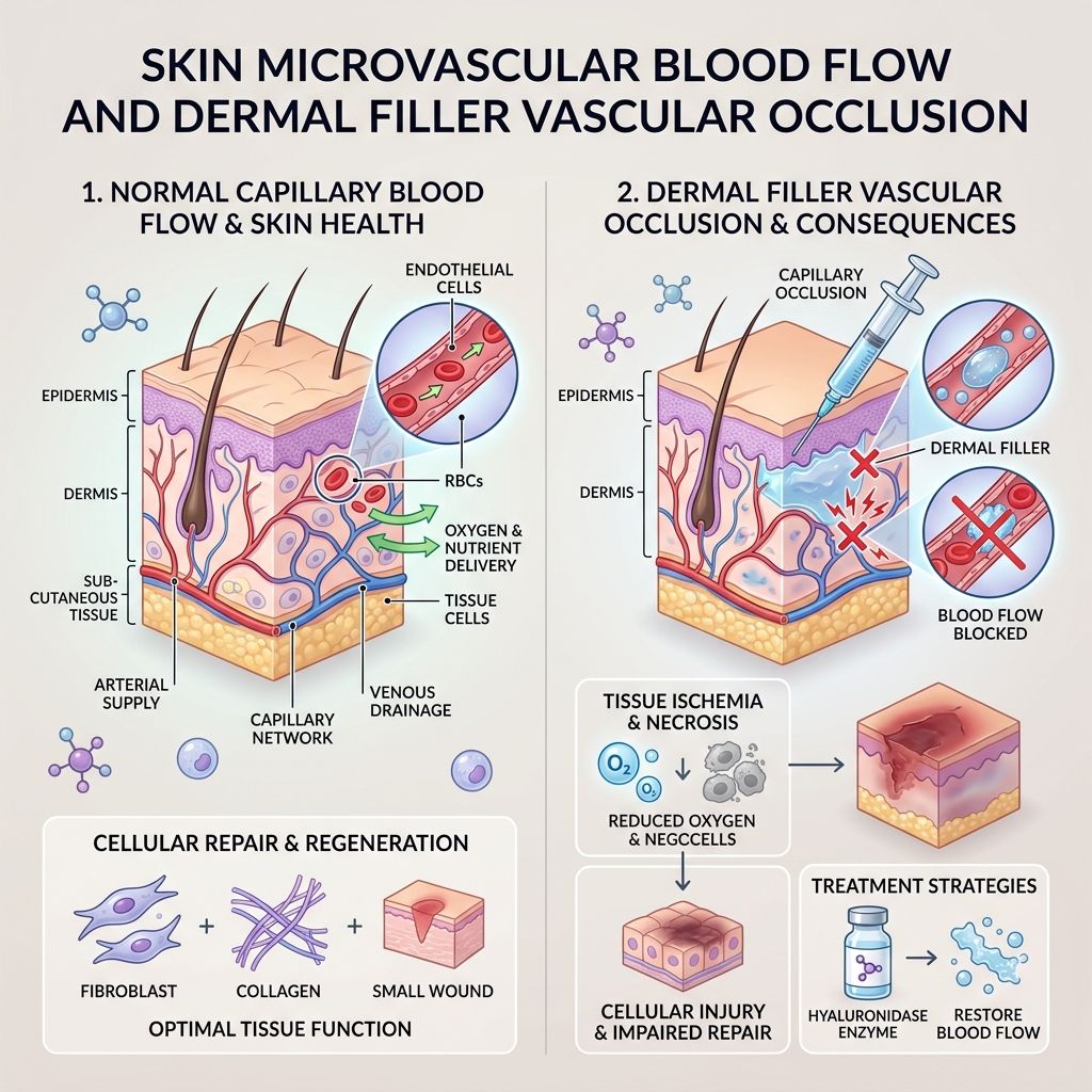
When injectable dermal filler inadvertently compresses or embolizes an arterial branch, local microvascular perfusion drops precipitously. Deprived of oxygen and essential nutrients, the affected cutaneous area manifests pallor, livedo reticularis, localized pain, and progresses into sterile pustules, exudate, and crust formation. Reversing this hypoxic cascade requires rapid reoxygenation to salvage ischemic tissue before permanent scarring occurs.
The 4-Part Combination Therapy Protocol
To ensure predictable, complete tissue recovery, Urgent Clinic implements a 4-part simultaneous treatment strategy:
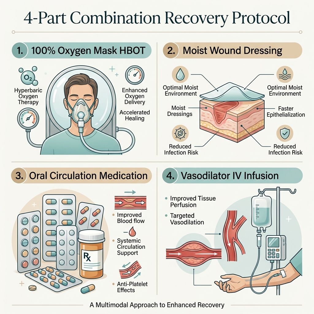
1. Hyperbaric Oxygen Therapy (HBOT): Administered in a hyperbaric chamber under elevated pressure for 60 to 90 minutes per session. Patients MUST wear a dedicated oxygen mask. Breathing chamber ambient air delivers only ~60% oxygen due to gas mixing, whereas mask-type delivery ensures 100% pure medical oxygen inhalation. Structured air breaks (hyperoxic-hypoxic paradox) trigger systemic stem cell mobilization and accelerate angiogenesis.
2. Specialized Wound Dressing: Regular removal of exudate and necrotic crusts, maintaining a controlled moist wound environment to prevent secondary bacterial infection and facilitate rapid re-epithelialization.
3. Oral Pharmacotherapy: Administration of antiplatelet and rheological agents to enhance microvascular blood flow, alongside targeted systemic antibiotics until exudate production ceases completely.
4. Intravenous (IV) Infusion Therapy: Co-infusion of hydration, high-dose Vitamin C antioxidants, and microvascular vasodilators (Niacin, Ginkgo biloba extract) to dilate constricted vessels, optimize cutaneous blood flow, and provide essential cofactors for collagen synthesis.
The 3-Stage Recovery Timeline & Treatment Milestones
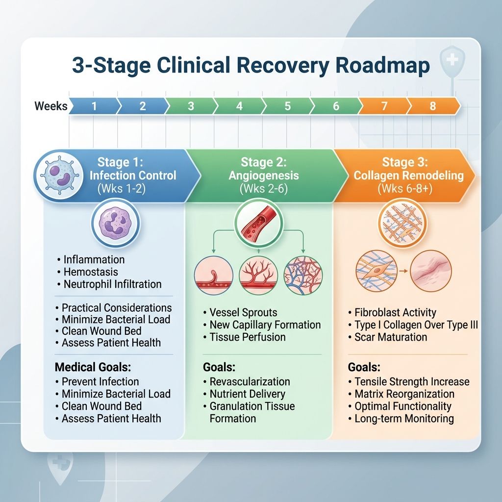
Under the 5-sessions-per-week protocol (60–90 min/session), patient recovery follows three distinct clinical stages:
• Stage 1 (Weeks 1–2, ~8–10 sessions): Infection Containment & Demarcation. Daily wound care, oral antibiotics, and intensive HBOT halt progressive tissue necrosis. Exudate diminishes and wound margins become clearly demarcated.
• Stage 2 (Weeks 2–6, Cumulative 20–30 sessions): Angiogenesis & Granulation. HBOT-stimulated growth factors induce microvascular capillary sprouting. Necrotic crusts naturally separate, revealing healthy pink granulation tissue underneath.
• Stage 3 (Weeks 6–8+, Cumulative 30–40 sessions): Re-epithelialization & Scar Management. The skin surface fully closes, and erythema gradually fades. Long-term scar and pigment remodeling continues over subsequent months.
Clinical Warning Signs & Home Care Guidelines
Patients must notify their medical team immediately if experiencing red-flag symptoms: sudden exacerbation of pain, swelling, or heat; foul-smelling purulent discharge; fever ≥38°C; or expanding dark necrotic boundaries. At home, patients must complete all prescribed oral medications, refrain from picking or peeling scabs, and strictly adhere to smoking cessation, as nicotine causes severe peripheral vasoconstriction that severely impairs healing.
Frequently Asked Questions (& Patient Guide)
Q: How many HBOT sessions are needed for filler necrosis recovery? A: Most clinical protocols require 20 to 40 sessions over 4 to 8 weeks, administered 5 days per week for 60 to 90 minutes per session, depending on necrosis depth and wound extent.
Q: Why is a mask mandatory during hyperbaric oxygen therapy? A: Breathing chamber air without a mask delivers only ~60% oxygen due to atmospheric gas mixing. A dedicated mask guarantees 100% pure medical oxygen inhalation and enables hyperoxic-hypoxic air breaks to maximize stem cell mobilization.
Clinical References & Scientific Evidence
1. DeLorenzi C. (2017). New High Dose Pulsed Hyaluronidase Protocol for the Treatment of Inadvertent Intra-Arterial Injection of Hyaluronic Acid Fillers. Aesthetic Surg J.
2. Aristizabal-Torres et al. Hyperbaric Oxygen Therapy in Aesthetic Complications. J Clin Aesthet Dermatol (JCAD).
3. Hadanny A, Efrati S. (2020). The Hyperoxic-Hypoxic Paradox. Biomolecules.
4. Kim DW et al. (2014). Wound Healing Algorithm for Filler-Induced Skin Necrosis. J Korean Med Sci.
Frequently Asked Questions
What are the early warning signs of vascular occlusion or skin necrosis after hyaluronic acid (HA) filler injections?
Early signs include sudden blanching (skin turning pale/white), severe localized throbbing pain exceeding normal post-injection tenderness, delayed capillary refill time (>2 seconds), and dusky purple mottling (livedo reticularis) within 24–48 hours.
How is filler vascular occlusion treated at a medical clinic in Seoul?
Urgent treatment involves immediate high-dose hyaluronidase enzyme injections to dissolve the arterial filler blockage, followed by warm compresses, oral vasodilators, hyperbaric oxygen therapy (HBOT) to restore oxygen supply to ischemic tissue, and sterile wound dressing for exudate management.
Can skin necrosis from filler complications be fully healed without scarring?
Yes, if treated within the early 24–72 hour window using emergency hyaluronidase, HBOT oxygenation, and medical-grade growth factors (EGF/PDRN). Prompt intervention prevents permanent scarring and tissue loss.
This article is for general information and is not medical advice. Consult a qualified healthcare provider about your individual situation.